Instrumentation
-
Shear Cells and Rheometers
-
Strain-controlled Rheometer
-
Light Scattering
-
Birefringence and Dynamic Light Scattering
-
Microscopes
-
General purpose equipment
Most of the experimental equipment, including specialized sample environments, is designed and built in-house. We give an overview of such equipment and sample cells, as well as the (partly) commercially available experimental set ups. The very last item is a list of available standard equipment.
Shear Cells and Rheometers
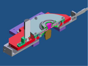
Shear Cell for MicroscopyWe have a set-up where we can perform microscopy in the zero-velocity plane, due to the fact that we can independently move the cone and the plate of our home-build shear cell. The plate is made from 0.17 mm sapphire windows, such that we can use high NA emersion objectives (up to NA=1.4). In the detection line we can either use a fast scanning confocal microscope (VTinfinity with a maximum frequency of 1000 frames per seconds, depending on the camera) or bright field (fluorescence). |
 |
Shear Cell for Light ScatteringThe setup allows for (time resolved) light scattering measurements under shear from a transparent Couette cell with rotating inner cylinder. We can go through none gap, if needed and project the scattered light is projected on a fast CCD or sCMOS camera. We probe structures between 250 nm and several microns. We can use the set-up also in combination with a microscope to perform microscopy under shear, using cells with a flattened surface. |
 |
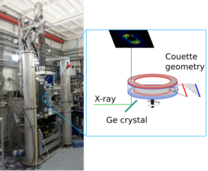
Vertical Rheo-SAXS Detection LineOur home-built vertical rheo-SAXS detection line allows us to combine all relevant geometries for rheology with small-angle X-ray scattering. Thus, we can probe all relevant scattering planes and scan the gap of the couette cell while having optimal rheology conditions. The detection line can be build up in synchrotron hutches that are high enough, as we did in ESRF and DIAMOND. |
 |
Strain-controlled Rheometer
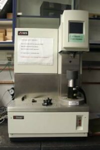
| TA Instruments ARES with Sealed Bath
Transducer: Tools: |
 |
Light Scattering
| Spatially Resolved Heterodyne Light Scattering
Velocity profiles along the gradient direction in an optical Couette cell can be probed. Both a back- and forward-scattering configuration with a difference in the scattering wave vector of about a factor of 10 can be used, for slow and fast decaying correlation functions. |
 |
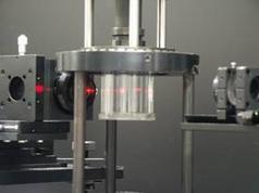
| Evanescent Dynamic Light Scattering
Our home built EWDLS instrument allows measuring the Brownian dynamics of colloids in the ultimate vicinity of a solid surface. The set-up is equipped with a 532 nm Nd-Yag Laser, two Perkin Elmer avalanche diodes and an ALV 6000 multiple tau correlator. Due to the triple axis geometry, it is possible to distinguish between motion parallel and normal to the wall. Besides the standard sample cell, a shear cell is available, which enables measuring particle velocities near the wall and local shear gradients. |
 EWDLS sample stage with shear cell. |
| Total Internal Reflection Microscopy
A home built TIRM set-up is available for the measurement of interaction profiles between a probe particle and a flat transparent surface. Forces can be measured down to the femto-Newton range. For standard measurements a photomultiplier is used to measure scattered intensity traces. To measure interaction profiles under shear, a specialized flow-through cell and a high-resolution CCD camera can be applied. |
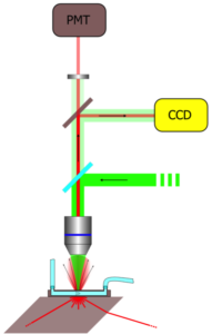 Sketch of the set-up for total internal reflection microscopy. |
| Thermal Diffusion Forced Rayleigh Scattering (TDFRS)
A grating created by the interference of two near infrared laser beams (λ=980 nm) is written in a sample. A weak absorption band in the aqueous solution, converts the intensity grating into a temperature grating, which in turn causes a concentration grating by the effect of thermal diffusion. Both gratings contribute to a combined refractive index grating, which is read out by diffraction of a third laser beam. Analyzing the time dependent diffraction efficiency, the three transport coefficients can be obtained. The thermal diffusivity Dth, the translational diffusion coefficient D, and the thermal diffusion coefficient DT. The ratio of the thermal diffusion coefficient and the translational diffusion coefficient allows the determination of the Soret coefficient ST. |
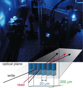 TDFRS-experiment with a principal sketch. |
Birefringence and Dynamic Light Scattering (DLS) in External Fields
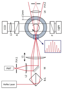
A null-birefringence set-up with an oblique incident laser beam (as shown in Fig.1) enables probing the birefringence of samples with an anisotropy of the refractive index that is perpendicular to the walls of the sample cell. An example is the alignment of rod-like colloids along the electric-field direction that is generated by two transparent electrodes (shown in the sketch in Fig.1).
This cell consists of two ITO plates, separated by a Teflon spacer, which are connected to an AC-voltage generator. This cell can also be used in a microscope, as well as a vertical DLS set-up shown in Fig.2. like the birefringence set up, the DLS set-up uses a He-Ne laser with a 10 mW intensity. The angular range of the vertically aligned DSL set-up is a few degrees to about 70 degrees [1].
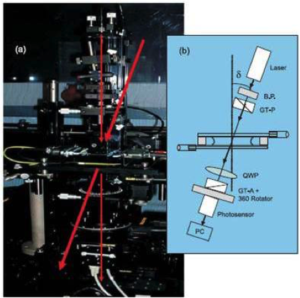 Fig.1: The birefringence set-up |
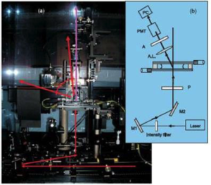 Fig.2: The DLS set-up
|
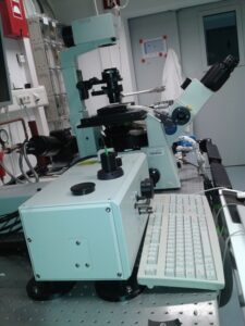
Microscopes
Microscopy is crucial for our research and is used to study the evolution of mesoscopic structures in external fields down to the single particle level. Therefore, our microscopy set ups include microscopes ranging from ultra-fast light microscopy to dedicated transition electron microscopy.
|
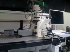
Light Microscope
Cameras: Andor iXon-Ultra-888 (1024 x 1024 pixels, 26 fps); Andor sCMOS (2560 x 2160 pixels, 100 fps); Hamamatsu C9100 EM-CCD (1000 x 1000 pixeks, 30 fps) and others. |
|
|
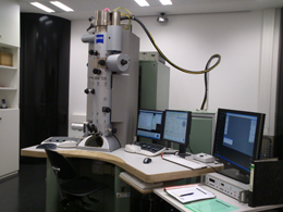
Transemission Electron Microscope (TEM) Our laboratory is also equipped with a Zeiss Libra 120 Transmission Electron Microscope (TEM), operating at 120 keV. It uses a LaB6 cathode, has an in-column energy filter, can be operated in STEM mode and is equipped with a Gatan US1000 (2k x 2k) CCD camera. The microscope is used for routine characterization of silica and gibbsite-based model systems, for example, and in the development of novel colloid systems. Working in cooperation, other groups also make use of the microscope, e.g. in the study of protein aggregation. |
 |
General purpose equipment
|
Experimental Equipment |
Synthesis Equipment |
|
|
|
|
|
|
|
|
|
|
|
|
|
|
|
|
|